Role of Electrophysiologic Study (EPS)
What is an electrophysiologic study (EPS)?
An electrophysiologic study (EPS) is used to find the source of abnormal heart rhythms. Some of these rhythms are caused by abnormalities in the heart’s conduction system. The conduction system is made up of special cells linked together in pathways to carry the electrical impulse that causes the heart to beat and pump.
EPS tests the condition of your heart’s conduction system by measuring the speed of an electrical impulse traveling through the system. It locates conduction pathways that are in the wrong place. EPS helps to identify many types of abnormal heart rhythms by trying to reproduce them.
Who needs an EPS?
Most people with abnormal heart rhythms don’t need EPS. The proper diagnosis is usually made by an electrocardiogram.
EPS is useful if you have collapsed suddenly and your doctor can’t find a good reason for the incident. It is especially useful if you have been revived after a cardiac arrest and there is no evidence of heart damage. Without treatment, there is a good chance of your having another collapse from these problems.
A few people have abnormalities of the conduction pathways of the heart. The normal pathway may be altered in such a way that very fast heart rates and even death result. EPS helps to locate the position of these abnormal pathways so that treatment can be started.
EPS may also be used to test the effect of various treatments, usually medicines, on the heart rhythm abnormality. Because the abnormal rhythm often can be reproduced by EPS, different medicines can be tested to see how well they work.
How is an EPS done?
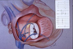
The EPS procedure may last an hour or more. It is rarely painful. You will be given drugs to help you relax, but you will remain awake during the procedure. Small wires called electrode catheters are put into your bloodstream through a vein. A drug is injected into your skin around the vein to prevent pain. Several catheters may be inserted at the same time. The catheters are put into positions in the heart’s right upper chamber (right atrium) and right lower chamber (right ventricle). The positions are checked by x-ray.
The outside end of the catheter is attached to a recorder that can measure the speed of electrical impulses inside your heart. By making different measurements, the doctor can locate your heartbeat conduction pathways and assess their condition. Sometimes an attempt must be made to reproduce the abnormal heart rhythm. The outside end of a catheter is attached to a device that sends an electrical charge through the catheter. The charge may be able to reproduce the abnormal rhythm you had. If so, your doctor can identify the problem and suggest the best treatment for you.
When the test is over, the catheters are removed. Pressure is applied over the veins until bleeding stops. In most cases, you can get up and move about after 3 to 6 hours. If a life-threatening problem is not identified, you probably will not need to stay in the hospital overnight.
Is EPS dangerous?
There is risk with every treatment or procedure. Talk to your Cardiologist for complete information about how the risks apply to you. Problems rarely occur. Most of these problems are minor, such as a little bleeding around where the catheters were inserted. The doctors and medical team who do the procedure are trained to manage any problems that may come up. A heart rhythm problem sometimes starts during EPS that needs immediate treatment. This treatment sometimes requires the delivery of an electric shock across the chest. This treatment, called defibrillation, restores the heart rhythm to normal. You will not feel any discomfort from the shock.